
%402x.svg)

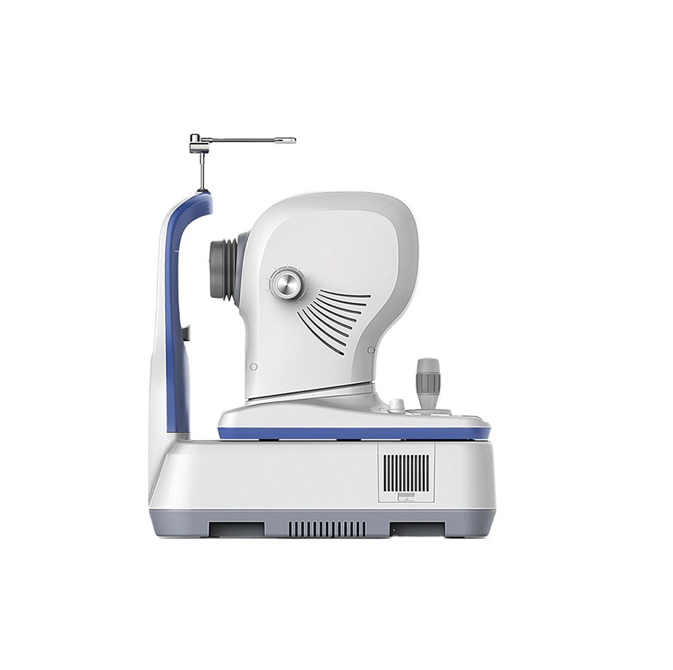
OSE-3000 Optical Coherence Tomography
.png)
Description
OSE-3000 Optical Coherence Tomography Features
The OSE-3000 Optical Coherence Tomography (OCT) system offers a range of advanced features for comprehensive eye examination and analysis.
MACULA Imaging:
- LSO (Line Scanning Ophthalmoscopy): Equipped with LSO, the MOcean 3000 provides high-quality fundus imaging. This aids physicians in localizing lesions effectively.
- Macula HD Line: High-definition OCT imaging unveils small lesions. Switchable OCT scan lengths between 6mm and 12mm.
- Macula Six-line Radial: Provides a quick retina overview with HD imaging and rapid data analysis.
- Software Analysis: Includes retinal thickness analysis, ganglion cell analysis, and high-definition OCT imaging with 5-image averaging.
- Macula Cube: Point-by-point retinal thickness assessment using a dense 500 x 100 cube.
- Software Analysis: In-depth features such as retinal thickness analysis, progression analysis, 3D visualization, and en-face analysis.
GLAUCOMA Analysis:
- Comprehensive glaucoma analysis with two scan modes: glaucoma cube scan in macular and disc areas.
- Software Analysis (Macular): Ganglion cell analysis, progression analysis.
- Software Analysis (Disc): RNFL analysis, cup-disk analysis, calculation circle and circle scan tomogram, progression analysis, OU comparative analysis.
- Informative Report: Offers OU comparative analysis and progression analysis report.
ANTERIOR SEGMENT Assessment:
- Anterior HD Line: High-definition OCT imaging of the cornea, enabling localization of Bowman’s layer, Anterior Chamber Angle, and manual measurements.
- Anterior Six-line Radial: Measures central corneal thickness through 6 radial lines. Software analysis includes corneal pachymetry and manual measurements.
PREMIUM FUNCTIONS:
- En-face Analysis: Enables precise lesion localization within subretinal layers.
- Choroid View
- IS/OS-Ellipsoid View
- Mid-Retina View
- VRI View
- 3D En-face View
Network System: Remote Analysis System:
- Moptim: Provides remote viewer software for displaying, enhancing, analyzing, and saving images from MOcean 3000/3000plus.
- Remote Diagnosis System: Specialists at Big Picture Eye Health review customer scans remotely for over 45 eye pathologies. Generates instant reports, including educational content and specialist referrals.
The OSE-3000 OCT system offers a comprehensive suite of features, from macula and glaucoma analysis to anterior segment assessment and premium functions for in-depth analysis. The integrated network system facilitates remote analysis and diagnosis for enhanced patient care.
Product Dimensions
Width
cm
Height
cm
length
cm
Weight
g
Product Specifications
Specifications
Methodology
Spectral Domain Oct
Optical Source
Super luminescent diode (SLD), 840nm
Scan speed
36000 A-Scan/s
Axial Resolution(optical)
5 microns (optical) 2.7 microns (digital)
Tranverse resolution
15 microns (optional), 3 microns (digital)
A-Scan depth
2.3MM
Diopter range
-20D to +20D
Scan patterns
Macular :HD line scan (6mm or 12mm),3D scan (6mm*6mm) Anterior:HD Line scan (6mm), 6-line radial scan
FUNDUS IMAGING
Methodology
Line scanning opthalmoscopoy (LSO)
Frame rate
10fps
Minmum pupil diameter
3.0mm
Field of view
47 degrees
SOFTWARE ANALYSIS
Macula
Retina thickness analysis,3D view, En-face analysis, Progression analysi
Glaucoma
RNFL analysis, Ganglion cell analysis , Cup-disk analysis,progression analysis ,OU comparative analysis.
Anterior segment
Manual measurement, Corneal thickness analysis
Others
DICOM conformance,Remote viewer software available
ELECTRICAL AND PHYSICAL
Weight
29kg
Dimension
450mm(l)*250mm(W)*450MM(H)
Source voltage
AC 100-240V
Frequency
50Hz-60Hz
Power input
90VA
Shipping
We offer country-wide shipping.
Return & Exchange
If you are not satisfied with your purchase you can return it to us within 14 days for an exchange or refund. For more info email us at info@internationalopticalindustries.com
Help
Email us at info@internationalopticalindustries.com
About International Optical Industries

Welcome to International Optical Industries - the hub of innovative ophthalmic solutions, headquartered in the vibrant city of Miami, Florida. Since our establishment in 2006, we have taken pride in providing the international market with high-quality, state-of-the-art ophthalmological equipment. Over looked by a team assuring product that aligns at best price and quality with on sight technicians and learning opportunities on operation


.png)